Δευτέρα 2 Δεκεμβρίου 2013

Οστεοπαθητική και Θηλασμός (κολικοί- Βοήθεια στο βρέφος και τη μαμά)

Ανατομία του Βρεφικού κρανίου
μετωπιαίο οστό
βρεγματικά οστα
κροταφικα οστα
σφηνοειδη οστα
ινιακό οστό
Fontanelles


Γιατί όχι πάντα Καισαρική;
ΤΟ ΜΩΡΟ ΠΡΕΠΕΙ ΝΑ ΠΕΡΑΣΕΙ ΑΠΟ ΤΟ ΓΕΝΝΗΤΙΚΟ ΚΑΝΑΛΙ 
Με τις πιέσεις : 
Βγαίνει υγρό απο τους πνεύμονες 
Δημιουργείται πίεση για την πρώτη δυνατή ΑΝΑΣΑ 
Διεγείρεται το κεντρικό νευρικό σύστημα και αρχίζει να οργανώνεται 
Αλλάζει δραματικά η λειτουργία του κυκλοφορικού 
Σταματαει η ομφαλιαία κυκλοφορία (σταματάει να πηγαινει και να έρχεται αίμα απο τον πλακούντα)
Κλεινει ωοειδες τρήμα (τρυπα επικοινωνιας αναμεσα στους καρδιακούς κόλπους)
Οι πνεύμονες φιλτράρουν και οξυγονώνουν το αίμα
Το συκώτι μεταβολίζει
Οι νεφροί φιλτράρουν αιμα
Το γαστρεντερικό σύστημα αρχίζει και απορροφάει θρεπτικά συστατικά
Οι αλλαγές λοιπόν που αναφέραμε πρεπει να γίνουν συστηματικά και με κάποια σειρά και ειναι πολύ απαιτητικές για το νεογέννητο
Οταν η μετάβαση  στην εξωμήτρια ζωή ειναι πολύ γρήγορη τότε όλες αυτές οι αλλαγές  γίνονται «ανοργάνωτα»
Οι οστεοπαθητικοί θεωρούν αυτές τις απότομες αλλαγές ως ένα σοκ ή μια ενόχληση η οποία «παραμένει» στο νευρικό σύστημα του μωρού.
Ετσι ενώ δεν υπάρχει εμφανές τραύμα . Το μωρό μπορεί να είναι εκνευρισμένο, γκρινιάρικο, ή αδύναμο.

 Θηλασμός
Ο θήλασμός είναι μια διαδικασία που για να γίνει σωστά απαιτεί συγχρονισμό μεταξύ μυών της γλώσσας, του φάρυγγα, του υοειδούς , της εμπρός αυχενικής περιοχής, και του διαφράγματος
 Η ηλεκτρομυογραφία δείχνει μεγαλύτερη λειτουργεια στους υπερυοειδείς μυες
Αιτίες που επηρεάζουν το θηλασμο 
Οι ικανότητες του μωρού να θηλάσει: δηλαδή να συγχρονισει, το άρπαγμα της θηλής, το ρούφηγμα, την κατάποση και την αναπνόη
Μητρικά προβλήματα: πληγωμένες θηλές, μαστίτιδες, πετρωμενα στήθη, μειωμενη παραγωγή (πολύ σπανιότερο το τελευταίο απο ότι νομιζει ο πολύς κόσμος)
Κοινωνικοί, πολιτισμικοί, προσωπικοί παράγοντες  
Η θέση της Οστεοπαθητικής 
 Πολλοί οστεοπαθητικοί πιστεύουν ότι οι δυσκολίες στο θηλασμό πολύ συχνά οφείλονται σε γενικότερες σωματικές δυσλειτουργίες , επηρεάζουν και αλλες συμπεριφορές του βρέφους (κολικούς ανησυχία, κλπ), και ξεκινούν από μικρότερα ή μεγαλύτερα τραύματα κατά τη γέννα
Άλλοι επικεντρώνονται στη σχέση τραύματος γέννας σε σχέση με τα οστά του κρανίου και την ανώτερη αυχενική σπονδυλική στήλη
 Κ.Ι.S.S 
Kinematic Imbalances due to Suboccipital Stress (Beidermann H ,Manual Therapy in Children 2004)
To ΚΙSS βασίζεται στην παρατήρηση συγκεκριμένης αναπτυξιακής ακολουθίας, κατα τα αλλα υγειών βρεφών
Χαρακτηρίζεται από μια αλληλουχία συμπτωμάτων: ανήσυχη συμπεριφορά, προβλήματα θηλασμού, κολικό, σε βρέφη ηλικίας μέχρι 2 μηνών.
Υποστηρίζεται ότι η δυσλειτουργία στην ανω αυχενική μοιρα που επηρεάζει την ιδιοδεκτηκότητα (η δυνατότητα που έχει το Κεντρικό Νευρικό Σύστημα να φέρνει σε επαφή και να συντονίζει τα διάφορα τμήματα του σώματος μεταξύ τους), η μειωμενη δυνατότητα να κατευθυνθει το κεφάλι προς το ερέθισμα και η μυική ένταση , ΟΛΑ επηρεάζουν την συμπεριφορά και την ανάπτυξη του βρέφους.
 Κολικός
Οι οστεοπαθητικοί υποστηρίζουν ότι μπορεί να οφείλεται και σε ερεθισμό του πνευμονογαστρικού νεύρου  το οποίο (βγαίνει απο τη βάση του κρανίου ανάμεσα στο ινιακό και το κροταφικό οστό) εχει πολλαπλες λειτουργίες: Νευρώνει τους μυς του λαρυγγα και του φαρυγγα, τα θωρακικα και κοιλιακα σπλάχνα μέχρι τη σπληνικη καμπή, αίσθηση της γεύσης από την επιγλωττιδα. Εξυπηρετεί την κατάποση και την φώνησης.
Η επίσημη εφημερίδα της αμερικάνικης παιδιατρικής ακαδημίας http://pediatrics.aappublications.org/content/early/2011/03/28/peds.2010-2098.full.pdf  αναφέρει ότι η οστεοπαθητική βοηθάει τον βρεφικό κολικό, και προτείνει να γίνουν περισσότερες έρευνες στο θέμα
Τί θα συμβεί όταν θα πάω το παιδί μου στον Οστεοπαθητικό 
Στην πρώτη μας επίσκεψη, θα πάρουμε ένα λεπτομερές ιστορικο του παιδιού με πληροφορίες για την εγκυμοσύνη, τη γεννα, την υγεία του παιδιού και των γονέων , την ψυχολογία του παιδιού και των γονέων.
Στη συνέχεια το μωρό θα εξεταστεί προσεκτικά για όλες τις εντάσεις για τις οποίες μιλήσαμε στην παρουσίαση.
Με πολύ απαλές κινήσεις και αγγίγματα ο οστεοπαθητικός θα προσπαθήσει να χαλαρώσει τις εντάσεις και να επαναφέρει την ισορροπία στο σωματάκι του μωρού 
Συνήθως μετά από 3-4 θεραπείες ο γονιός θα διαπιστώσει διαφορές και στον θηλασμό αλλά και στη διάθεση του μωρού (μπορεί να χαλαρώνει πιο εύκολα, να μη κλαίει τόσο όσο συνήθως, να κάνει κινήσεις με μεγαλύτερη ευκολία να έχει περισσότερη ενέργεια κ.α) 
 Μανούλες
Το συχνότερο σύμπτωμα κατά την εγκυμοσύνη και τους αμέσως επόμενους μήνες είναι σαφώς η οσφυαλγία, ωστόσο οι μυϊκοί πόνοι στην πλάτη, τον αυχένα και τους ώμους είναι επίσης αρκετά συχνοί. Ενδιαφέρον στοιχείο είναι ότι γυναίκες που πέρασαν την περίοδο της εγκυμοσύνης χωρίς ιδιαίτερα προβλήματα, μπορεί να παρουσιάσουν πόνους στην πλάτη και τη μέση στο διάστημα της λοχείας αλλά και μετά. Αυτό φαίνεται να έχει πολύ στενή σχέση με τον τρόπο που σηκώνουν και κρατούν το βρέφος τους καθώς και με τη στάση τους κατά το θηλασμό.

Τετάρτη 20 Νοεμβρίου 2013

Πυελικός Πόνος - Δυσλειτουργία της ηβικής σύμφυσης

Τι είναι η  δυσλειτουργία ηβικής σύμφυσης (SPD-Symphysis Pubis Dysfunction);

H SPD, γνωστή και ως Πυελικός Πόνος (PGP-Pelvic Girdle Pain), ορίζεται ως : ήπιος έως οξυς πόνος πάνω από την ηβική σύμφυση, ο οποίος μπορεί να εκτείνεται κάτω στην βουβωνική χώρα και στους μηρούς. Ορισμένες μελέτες αναφέρουν ότι έως και το 50% των εγκύων γυναικών έχουν κάποιο είδος της PGP πριν από τις 20 εβδομάδες κύησης.

Ο πόνος μπορεί να σχετίζεται με ένα προυπάρχον πρόβλημα που μέχρι τότε δεν έχει προκαλέσει πόνο, ή μπορεί να εμφανιστεί μετά από έναν τραυματισμό, συχνά ήσσονος σημασίας, κατά τη διάρκεια της εγκυμοσύνης ή του τοκετού.
Υπάρχουν παράγοντες που μπορεί να επιδεινώνουν την κατάσταση:
Η σταθερότητα του κορμού
Η αδυναμιά των μυών του πυελικού εδάφους
Παλαιότεοι τραυματισμοί
Προβλήματα της σπονδυλικής στήλης
Μυικές εντάσεις
Άλλες κατασκευαστικές Ανισσοροπίες

Τέτοιοι βασικοί παράγοντες σε συνδυασμό με την αύξηση του βάρους του μωρού που αναπτύσσεται και το μαλάκωμα των συνδέσμων επηρεάζουν τον ορθό καταμερισμό βάρους και την κίνηση μέσα στη λεκάνη.

Ποια είναι τα συμπτώματα;
Το κύριο σύμπτωμα της SPD είναι ο πόνος ή δυσφορία στην περιοχή της πυέλου, που συνήθως συγκεντρώνεται γύρω από την ηβική σύμφυση στο μπροστινό μέρος. Ο πόνος μπορεί να επεκταθεί και στις Ιερολαγόνιες αρθρώσεις ,στη βάση της σπονδυλικής στήλης, τη βουβωνική χώρα, τους μηρούς, τους γοφούς, κάτω κοιλιακή χώρα, και την πλάτη.
 
Η ένταση του πόνου της SPD μπορεί να ποικίλει. Μερικές γυναίκες αισθάνονται μόνο περιστασιακή ήπια δυσφορία, ενώ σε πιο ακραίες περιπτώσεις ο πόνος μπορεί να είναι εντονοτατος και να εμποδίζει τη φυσιολογική δραστηριότητα.
Τα συμπτώματα μπορεί να εμφανιστούν ανά πάσα στιγμή από το τέλος του πρώτου τριμήνου μέχρι ακόμη και μετά τη γέννηση.Συνήθως εμφανίζονται γύρω στα μέσα της εγκυμοσύνης.
Τα συμπτώματα αυτά επιδεινώνονται από οτιδήποτε ζορίζει οποιαδήποτε από τις αρθρώσεις της λεκάνης, ιδιαίτερα την ηβική σύμφυση. Για παράδειγμα:
• Η έγκυος στέκεται στο ένα πόδι για υπερβολικά μεγάλο χρονικό διάστημα - για να αλλάξει ρούχα
• Κινήσεις τέτοιες που απομακρύνουν τα γόνατα μεταξύ τους (και κατ' επέκταση ζορίζουν τα ισχία) - κάθισμα σταυροπόδι, πρόσθια κολύμβηση, είσοδος και έξοδος από το αυτοκινητο, οι περιστροφές στο κρεβάτι, το σεξ με πολύ ανοιχτά πόδια.
• Ιδιαίτερα έντονο περπάτημα με μεγάλους διασκελισμούς.

Πώς μπορεί να βοηθήσει η οστεοπαθητική;
Εάν μια ασθενής επισκεφθέι τον οστεοπαθητικό της αρκετά νωρίς δηλαδή μεταξύ 16-22 εβδομάδες, μπορεί να δει αρκετά μεγάλη ανακούφιση συνήθως μετά από 5-6 θεραπείες.

Ο οστεοπαθητικός μας μπορεί
• Να διορθώσει τις ανισορροπίες που υπάρχουν στην σπονδυλική στήλη, και τη λεκάνη (συνήθως προ-υπάρχουσες δυσλειτουργίες και ασυμμετρίες)
• Να απελευθερώσει από οποιαδήποτε ένταση στους μυς και τις αρθρώσεις
• Να παρέχει συμβουλές σχετικά με τη στάση του σώματος και την άσκηση
• Να παρέχει συμβουλες για την ενδυνάμωση του πυελικού εδάφους
 
Τι μπορεί να κάνει κάποιος που πάσχει από SPD ή από πόνο στη μέση για να βοηθηθεί κατά τη διάρκεια της εγκυμοσύνης;
 
Ο πυελικός πόνος και ο πόνος στην πλάτη και τη μέση δεν είναι φυσιολογικοί κατά την κύηση- αν και ειναι συνηθισμένοι. Ζητήστε βοήθεια το συντομότερο,μόλις ανακαλύψετε τον πόνο συμβουλευτείτε ένα καλό οστεοπαθητικό. Επιπρόσθετα:
 
• Αν κάτι πονάει, μην το κάνετε!
• Κοιμηθείτε με ένα μαξιλάρι ανάμεσα στα πόδια σας.
• Κρατήστε τα γόνατά σας κοντά  όταν γυρνάτε πάνω στο κρεβάτι.
• Κρατήστε τα γόνατά σας κοντά, όταν μπαινοβγαίνετε στο αυτοκίνητο - χρησιμοποιήστε μια πλαστική σακούλα πάνω στο κάθισμα για να σας βοηθήσει να περιστρέφεστε (αφαιρέσετε την πριν από την οδήγηση!)
• Ξεκουραστείτε όσο πιο συχνά μπορείτε
• Αποφεύγετε να κάθεστε σε σκληρές καρέκλες - Πάρτε ένα μαξιλάρι μαζί σας εάν χρειαστεί.
• Χρησιμοποιήστε μια μπάλα γέννας - μπαλα pilates για να κάθεστε.
• Προσπαθήστε να μην ανεβοκατεβαίνετε σκάλες.
• Μετρήστε την "ανώδυνη απόσταση-ανώδυνο κενό"  ανάμεσα στα γόνατά σας και ενημερώστε τη  μαία σαςκαι τον γυναικολόγο σας.
• Κάντε σεξ σε πλάγια θέση με υποστήριξη στα γόνατά σας.

Ποιες ασκήσεις πρέπει να αποφευγω;
• Βαθιά καθίσματα
• Lunges (εφορμήσεις)
• Κοιλιακούς τύπου situps
• Τρέξιμο
• Πρόσθια Κολύμβηση

Ποιες ασκήσεις να προτιμώ;
• Ασκήσεις ενδυνάμωσης του κορμού και του πυελικού εδάφους
• Ενίσχυση συγκεκριμένων κοιλιακών μυών με ειδικές ασκήσεις από ειδικό ώστε να μην επιβαρύνεται η κατάσταση 


Μετα τη γέννα;
Αν είχατε πυελικό πόνο κατά τη διάρκεια της εγκυμοσύνης, καλό είναι να επισκεφθείτε τον οστεοπαθητικό σας, το συντομότερο  μετά τη γέννα. Έτσι θα εξασφαλιστεί η ευθυγράμμιση της ηβικής σύμφυσης και η επιτάχυνση της ίασης. Επιπλέον θα λάβετε συγκεκριμένες συμβουλές και ειδικές ασκήσεις!
Σε πολλές γυναίκες ο πόνος και η δυσφορία που αισθάνθηκαν κατά τη διάρκεια της εγκυμοσύνης υποχωρεί μετά τη γέννηση. Ωστόσο, είναι σημαντικό τις πρώτες ημέρες και εβδομάδεςνα ξεκουράζονται όσο το δυνατόν περισσότερο, να αποφεύγουν τα περιττά βάρη και τους πολύ έντονους περιπατους.
Μετά από τις πρώτες εβδομάδες μπορείτε να αρχίσετε να κάνετε τις κατάλληλες ήπιες ασκήσεις για την ενίσχυση του κορμού, τους κοιλιακούς και το πυελικό έδαφος

Τετάρτη 17 Ιουλίου 2013

Οδηγηση - Οδηγίες για επιλογή σωστής θέσης, για να μειώσουμε τα μυοσκελετικά προβλήματα που σχετίζονται με την οδήγηση


ΟΔΗΓΗΣΗ

Η οδήγηση μπορεί να προκαλέσει:
Αυχεναλγίες, κεφαλαλγίες, οφθαλμαλγίες, πόνο στους (ώμους, αγκώνες, καρπούς, σπονδυλική στήλη, ισχίο, γοφούς, γόνατα, πόδια).

Πριν επιλέξουμε αυτοκίνητο 4 σημαντικά τεστ.

Το τέστ της προσευχής:
          Ενώνουμε τα 2 χέρια και τα τεντώνουμε ίσια μπροστά. Αν το τιμόνι είναι κεντραρισμένο τα χέρια μας πρέπει να έρθουν στη μέση του τιμονιού. Αν το τιμόνι δεν είναι στο κέντρο ο οδηγός τείνει να περιστρέφει το κορμί του για να ισορροπήσει τη θέση του τιμονιού με μακροπρόθεσμα προβλήματα στη μέση.

Το τεστ της γροθιάς:
Κάντε γροθιά με το δεξί χέρι και τοποθετείστε την πάνω από το κεφάλι. Θα πρέπει να χωράει άνετα ανάμεσα στο κεφάλι και την οροφή του αυτοκινήτου. Αν μόλις χωράει τότε ο χώρος είναι ανεπαρκής. Ο κίνδυνος μικρού χώρου κεφαλής είναι ο οδηγός να βουλιάζει στο κάθισμα για να ισορροπήσει τη διαφορά κάτι που ασκεί πίεση στη σπονδυλική στήλη και τους γοφούς. 

Το τέστ κάτοψης:
Με τα δύο χέρια στο τιμόνι κοιτάξτε τα πόδια σας. Θα πρέπει να μπορείτε να δείτε το ίδιο και τα δύο πόδια. Συχνά το ένα πόδι φαίνεται περισσότερο από το άλλο γιατί υπάρχει περιστροφή του ώμου σε σχέση με τη λεκάνη.

Το τέστ του Δεξιού ποδιού.
Αφού οδηγήσουμε για λίγη ώρα κοιτάμε κάτω το δεξί μας πόδι. Είναι πιο ψηλά από το Αριστερό ή έχει πέσει προς την άκρη της θέσης;. Το πόδι μας είναι περίπου σε ευθεία με το μηρό ή έχει έρθει προς το κέντρο του αυτοκινήτου.

10 σημαντικά σημεία κατά την οδήγηση:
1.     Η θέση πρέπει να βρίσκεται στις 95 με 100 μοίρες.
2.     Το προσκέφαλο πρέπει να βρίσκεται σε τέτοια θέση ώστε το κέντρο του να βρίσκεται στο ύψος των ματιών. Αν είναι πολύ χαμηλά μπορεί να αποβεί μοιραίο σε ένα ατύχημα
3.     Όταν μπαίνετε στο αμάξι, πρώτα καθίστε και μετά βάλτε τα πόδια μέσα. Όταν βγαίνετε, τραβήξτε τη θέση πίσω, βγάλτε τα πόδια και μετά το υπόλοιπο σώμα.
4.     Όταν οδηγείτε μην έχετε το Αρ πόδι πάνω από τον συμπλέκτη. Ξεκουράστε το στην ειδική θέση.
5.     Σε κάθε φανάρι, για να χαλαρώσετε, εισπνεύσετε υψώνοντας τους ώμους στα αυτιά, εκπνεύσατε χαλαρώνοντας τους ώμους.
6.     Αποφεύγετε να πιάνετε αντικείμενα από το πίσω κάθισμα. Μην τεμπελιάζετε. Βγείτε έξω και πάρτε το.
7.     Προσέχετε όταν γεμίζετε και αδειάζετε το αμάξι
8.     Αποφύγετε να κουβαλάτε βαριά αντικείμενα. Ζητείστε βοήθεια για να αλλάξετε ελαστικό
9.     Καθίστε με τα χέρια ελαφρώς λυγισμένα στους αγκώνες μη σκύβετε και μη φεύγετε μπροστά στη θέση
10.                        Φοράτε πάντα ζώνη η οποία να εφαρμόζει σωστά. Βεβαιωθείτε ότι τα παιδιά φορούν ζώνη και έχουν τα απαραίτητα μαξιλάρια.   

Δευτέρα 8 Ιουλίου 2013

Σκολίωση - Η οστεοπαθητική Αντιμετώπιση


Με τον όρο σκολίωση εννοούμε τη στρέβλωση της δομής του σώματος με τη δημιουργία καμπύλης προς μια κατεύθυνση. Συνηθέστερα εμφανίζεται στη σπονδυλική στήλη, αλλά μπορούμε να τη βρούμε στη λεκάνη, αλλά και στα οστά του κρανίου. Ετυμολογικά προέρχεται από την αρχαία ελληνική λέξη σκολιός που σημαίνει στραβός, στρεβλός (άρα ουδεμία σχέση έχει η σκολίωση με το σχολείο ).
 
Η σπονδυλική σκολίωση μπορεί να είναι κατασκευαστική, εξαιτίας κάποιου οστέινου ελαττώματος , π.χ απουσία ενός πλευρού ή ατελής ανάπτυξη ενός σπονδύλου. Μπορεί να είναι αποτέλεσμα νευρομυϊκής ασθένειας, όπως ειναι η εγκεφαλική παράλυση όπου οι μυες της σπονδυλικής στήλης εχουν μεγαλύτερη σπαστικότητα από τη μια πλευρά, ή και άλλων παραλυτικών καταστάσεων όπου οι μυες ειναι πολύ πιο αδύναμοι από τη μια πλευρά. Σε αυτές τις καταστάσεις οι μυες της σπονδυλικής στήλης ασκούν μεγαλύτερη δύναμηστην κάμψη προς τημια μερία σε σχέση με την άλλη. Χειρουργία κατάτη βρεφική ή παιδική ηλικία , μπορεί να δημιουργήσουν ουλές στη μια μεριά του κοιλιακού τοιχώματος, οι οποίες , κατά την ανάπτυξη, μπορεί να δημιουργήσουν καμπύλη στη σπονδυλική στήλη, επειδή οι μαλακοί ιστοί γύρω από την ουλή, μετατρέπονται σε ουλώδη και ινώδη ιστό και δεν έχουν την ιδια ελαστικότητα με την άλλη πλευρά.

Αυτές οι αιτίες σκολίωσης είναι σπάνιες. Πολύ συχνότερες είναι οι ιδιοπαθητικές εφηβικές λειτουργικές σκολιώσεις. «Ιδιοπαθητικές» γιατί δε γνωρίζουμε την αιτία, «εφηβικές» γιατί εμφανίζονται συχνότερα όταν ξεκινάει η εφηβεία και «λειτουργικές» γιατί δεν υπάρχει κατασκευαστική δυσμορφία

Υπαρχουν ωστόσο οριμένοι αιτιολογικοί παράγοντες που αναγνωρίζονται από τον οστεοπαθητικό και οι οποίοι αντιδρούν θετικά στην οστεοπαθητική θεραπεία. Για να τους εκτιμήσουμε , πρέπει να δεχτούμε τον ασθενή ως μια δυναμική λειτουργική μονάδα από την κορυφή ως τα νύχια και όχι απλά ως μια σπονδυλική στήλη που έχει μια ασυνήθιστη καμπύλη. Εξετάζοντας τον ασθενή όρθιο, ελέγχουμε το επίπεδο των αυτιών, τους ώμους, τις ωμοπλάτες, και τις λαγόνες για συμμετρία. Ελέγχουμε το πώς κατανέμει ο ασθενής το βάρος στα δύο του πόδια. Ελέγχουμε το πώς πατάει τα πέλματα. Ελέγχουμε αν η καμπύλη διορθώνεται όταν ο ασθενής σκύψει μπροστά. Ελέγχουμε την αντίδραση της καμπύλης όταν ο ασθενής λυγίζει το ένα ή το άλλο γόνατο. Ελέγχουμε τη συμμετρία στην κίνηση των πλευρών. Ελέγχουμε την ελευθερία της κίνησης των χεριών δίπλα στους ώμους.
Παραμένει η σκολίωση όταν ο ασθενής κάθεται; Εχει η λεκάνη ισορροπία; Έχουν τα πόδια το ίδιο μήκος; Έχουν την ίδια ένταση οι μύες της σπονδυλικής στήλης;
Στην συνέχεια ψιλαφούμε τον μηχανισμό των κρανιακών οστών για στρέβλωση θέσης ή ασυμμετρία κίνησης. Λειτουργικά, το σώμα μας κρέμεται από το κεφάλι, και έτσι, στρέβλωση  στον κρανιακό μηχανισμό από μια τραυματική γέννα, προδιαθέτει για δημιουργία σπονδυλικής σκολίωσης, καθώς η περιτονία μπορεί να έχει ασύμμετρη «ένταση» στο σώμα. Ορθοδοντικές παρεμβάσεις για να προκαλέσουν εξισσορόπιση στο σαγόνι, μπορεί να δημιουργήσει ή να επιδεινώσει τη σπονδυλική σκολίωση.

Για την κλινική εκτίμηση μπορεί να χρειαστεί μια όρθια ακτινογραφία όπου μπορούμε να εξετάσουμε όχι μόνο τη φύση και τις μοίρες της καμπύλης, αλλά και το μήκος των ποδιών

Η θεραπεία μπορεί να περιλαμβάνει οστεοπαθητικούς χειρισμούς στη λεκάνη, τον αυχένα, το κρανίο, τα πλευρά, την κοιλιακή χώρα, την περιτονία, καθώς και την περιοχή της εμφάνισης της σκολίωσης. Αν υπάρχει ανατομική ανισοσκελία (διαφορά μήκους των ποδιών) ίσως χρειαστούν πάτοι. Επιπλέον μπορεί να δοθούν συγκεκριμένες ασκήσεις οι οποίες θα επιταγχύνουν το αποτέλεσμα της οστεοπαθητικής θεραπείας. Προσοχή οι ασκήσεις δεν αντικαθιστούν την οστεοπαθητική απλά βοηθούν τον οστεοπαθητικό να έχει πιο γρήγορα αποτελέσματα.

Η τσάντα ή το σακίδιο πρέπει να ελέγχεται προσεκτικά. Δεν πρέπει να ειναι πολύ βαριά και θα πρέπει να ειναι ισορροπημένα και στους δύο ώμους .

Η χρήση κηδεμόνα μπορεί να ενδείκνυται για σοβαρές κατασκευαστικές σκολιώσεις. Το χειρουργίο μπορεί να χρειαστεί αν η σκολίωση έχει επιδεινωθεί γρήγορα (αύξηση μοιρών) ή αν υπάρχουν δομικές ανωμαλίες. Η εμπειρία , μας δειχνει ότι εάν προηγηθεί η οστεοπαθητική θεραπεία, αυτά τα πιο δραστικά μέτρα, χρειάζονται σπάνια. 


Ελευθέριος Μουράτης
Οστεοπαθητικός Φυσιοπαθολόγος
www.facebook.com/pages/Κέντρο-Οστεοπαθητικής-Φυσιοπαθολογίας-Ελευθέριος-Μουράτης/408655552499595

Τετάρτη 26 Ιουνίου 2013

Σύνδρομο υπερχρησης- Τραυματισμοί χρηστών Η/Υ με Βιοανάδραση - Πρωτοκολλο Εξέτασης και εκπαίδευσης - Αγγλική Γλώσσα

http://www.bfe.org/protocol/pro09eng1.htm


Introduction
Improper work habits, poor workstation ergonomics, and environment can lead to physiological dysregulation such as muscle soreness, fatigue, and injury (Grandjean, 1987). Some workers develop chronic neck and upper limb pain also known as repetitive strain injury. (RSI), cumulative trauma disorder (CTD) or overuse syndrome, from long hours of repetitive tasks at personal computer workstations. Workers with RSI suffer loss of productivity and income with increasing medical costs. RSI accounted for forty percent of workers compensation cases in 1990. Discomfort and injury can shape the way PC users feel about their job and computers. Forty million Americans use computers, and 15-20 percent are at risk for RSI symptoms (CDC, 1984). RSI threatens to inflict individuals with illness and overwhelm corporations with increasing medical costs and lost productivity.
At present, workstation ergonomic analysis, proper positioning of furniture and equipment, different mice and keyboards, and periodic rest may help reduce physiological dysregulation (the incidence of muscle fatigue or injury). This mechanical approach, however, lacks two crucial elements:
  • Kinesthetic awareness of psychophysiology by computer users.
  • Development of skills to inhibit inappropriate and excessive bracing during task performance.
The computer user must learn to reduce tension and relax muscles when they are not used for the task. Preventing RSI involves a combination of proper ergonomics, work pattern variation (work/rest cycle), and self-regulation through biofeedback to reduce dysponetic activity (inappropriate and misdirected, as well as unconscious muscle bracing). Without kinesthetic awareness and without the skills to reduce tension, ergonomic adjustments with intermittent rest periods are NOT sufficient to reduce risk for injury.
Computer users can learn preventative skills to sense muscle tension and incorporate relaxation and regeneration of muscles during data entry and mouse use. Biofeedback instruments can be used to monitor specific muscle sites and to warn the user of excessive strain or overuse habits that can lead to chronic pain or injury. This mastery process reduces the risk of RSI.
RISK FACTOR ANALYSIS
The development of RSI involves ergonomic and psychophysiological contributing factors which include:
  • Inappropriate ergonomic workstation setup
  • VDT (monitor) induced near-vision stress
  • Asymmetrical task performance
  • Restricted body movements
  • Absence of brief (1-2 seconds) regenerative breaks during work activity (micro-breaks)
  • Dysponesis during task performance (co-contraction, lack of inhibition of antagonist during movement)
  • Lack of somatic awareness of tension and relaxation
  • Physiological tension during self-perceived relaxation
  • Excessive focus on tasks and or flawless work record
  • Work dissatisfaction
  • Frequent previous illnesses
  • Excessive physiological reactivity
  • Thoracic breathing and/or breath holding during data entry
  • Presence of tender trigger points
The following RSI protocol includes ergonomic and work style evaluation, psychophysiological profile, risk factor analysis, biofeedback training and education.
ERGONOMIC AND WORK STYLE EVALUATION
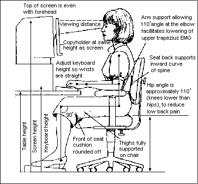
Ergonomic evaluation needs to be done at the work site under normal working conditions. (Compare the computer user position to the recommended guidelines, see Fig. 1.) The attached assessment form, SFSU WORKSTATION AND ERGONOMIC ASSESSMENT (Peper and Tibbetts, 1994) can be used as a guideline to cover most of the ergonomic risks (see also Jones, 1991; State of California, 1993). At the same time, movement analysis of task performance needs to be included because repeated asymmetrical movements increase risks (Donaldson, 1994). A table of typical computer user complaints, corresponding ergonomic factors, and areas for surface electromyography (sEMG) are listed in Table 1 (Wilson, 1994).

Figure 1. Workstation arrangement to reduce risk of discomfort. (Illustration, courtesy of Great Performance, Inc. (800)433-3803).
After evaluation, ergonomic risks are identified and solutions proposed. Many solutions can be economical, such as lowering the monitor by placing the computer sideways on the floor, lowering the keyboard by attaching a keyboard drawer, raising the feet by using telephone books, and removing boxes or waste basket from under the desk so that the legs are free to move. Sometimes, the solution requires a reorganization of the office, such as moving office furniture, to reduce excessive reaching, or to allow distant vision.
In cases of asymmetrical job patterns, the person is guided to do the job alternately using opposite sides of the body, as well as changing the position of furniture (phones, files) to balance movement patterns. For example, during the morning, place the phone on the left side and lift the receiver with the left hand, while during the afternoon, place the phone on the right side and use the right hand.
An additional factor commonly overlooked is appropriate vision correction, especially for people who wear bifocals. Prescribed reading glasses (often used at the computer) force the user to tilt the head back or hunch forward since the focal distance is not set at the distance of their workstation monitor (Grandjean 1987). Some users may need to have special computer monitor reading glasses.
Equally important, computer users need to be aware of the work/rest cycle. This means both very short breaks and movements during task performance and larger body movements during frequent longer breaks.
PSYCHOPHYSIOLOGICAL PROFILE
After ergonomic assessment and adjustments, a physiological assessment should be done. If possible, it should include an actual work site assessment, which can easily be done with a laptop computer and a portable two channel EMG device like Thought Technology's MyoTrac2(TM) or ProComp+(TM) systems. The comprehensive profile consists of different phases which can also be selectively used depending upon the computer user's needs.
The psychophysiological profile consists of physiological monitoring during: a) task performance, b) simulated emotional stress, c) extended data entry, and d) movement symmetry. The purpose is to assess dysponetic activity (misdirected and inappropriate bracing patterns), the length of short breaks, the effect of emotional stress upon physiological reactivity, physiological recovery, asymmetrical muscle co-contractions, and somatic awareness.
The profile assesses the individual at the computer, either at the job site or at a simulation of the workplace setting. The data entry task needs to include the actual data entry pattern of the user (e.g. keyboard, mouse, and/or trackball). The initial assessment does not include feedback, although all the data is recorded for later analysis.
IMPORTANT: The protocol should be adapted to assess real task performance such as job related data entry, reaching for the phone with the right and left hand, etc.
After the protocol has been completed, the data can then be reviewed with the client to show the individual's physiological responses. This information is then used to determine self-regulation strategies to improve health. This feedback is usually done directly after the assessment so that the information can be used to begin retraining.
SENSOR REQUIREMENTS AND PLACEMENT
The minimum requirement for the assessment is two channels of surface electromyography (sEMG). A two channel sEMG assessment will require moving sensor leads to different muscle sites during different phases of the assessment. A more comprehensive assessment would include a minimum of 4 channels of sEMG, respiration, skin temperature (Temp) and skin conductance response (SCR).

Two channel sEMG sensor placement:

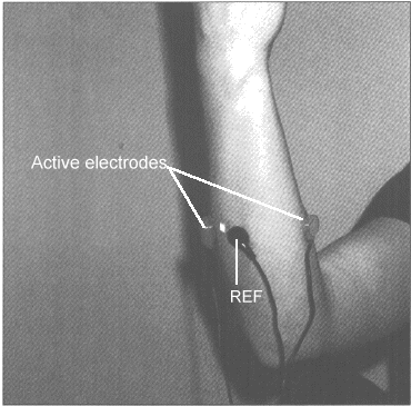
A) Forearm sEMG: Place the active electrodes midpoint on the extensor and flexor muscles (see Fig. 2) to monitor forearm tension. The purpose for monitoring the forearm is that most subjects do not relax the fingers or wrist muscles as long as the fingers are on the keyboard or holding the mouse.


Figure 2. Forearm sEMG sensor placement
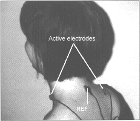
B) Neck and shoulders sEMG: Place one active electrode over the left scalene and the other, midpoint on the right trapezius (see Fig. 3). The purpose of monitoring the neck and shoulders is that most subjects raise their shoulders and tend toward thoracic breathing patterns during task performance. The left scalene to right trapezius placement will also monitor bracing by the scalene and sternocleidomastoid muscles.

Figure 3. Neck and shoulder sEMG placement

Four channel sEMG sensor placement:

Sensors are placed to allow bilateral analysis during the symmetry assessment. The most common bilateral electrode placements (right and left) include the following muscles: upper trapezius, lower trapezius, sternocleidomastoid, rhomboid, and pectoralis. The purpose for monitoring upper and lower trapezius is to enhance scapular stabilization since the lower trapezius will inhibit the upper trapezius activity. (For exact electrode locations see: Soderberg, 1992: This manual is free and available from NIOSH, see recommended sources for more information; and Basmajian & Blumenstein, 1980).
Comprehensive monitoring includes thoracic and abdominal respiratory patterns and breathing rate, peripheral temperature, skin conductance, and heart rate. Monitoring should also include the following sEMG placements, bilateral cervical paraspinals, masseters, temporalis, deltoid, upper and lower trapezius, pectoralis, infraspinatis, wrist extensors and flexors, and possibly, tibialis and gastrocnemius.

ASSESSMENT PROTOCOL
  • Describe the protocol sequence.
  • Have the person sit comfortably at the computer station. (Do the ergonomic assessment and modifications as needed)
  • Attach physiological monitoring device and verify signals.
  • After each step of the assessment, have the person rate their subjective stress/tension.
PHASE 1:
Effect of Position and Task on Physiology
PURPOSE:
To assess subjective muscle tension awareness, posture and task performance upon the physiology.

PROCEDURE:
  1. Sit comfortably with hands resting on lap (30 seconds baseline).
  2. Place fingers comfortably (their normal mode) on the keyboard at home row (30 seconds).
  3. Type a standard text (a sample letter or materials that simulate a normal job task) (60 seconds).
  4. Place fingers comfortably on the keyboard without pressing keys (30 seconds).
  5. Place hands back on lap comfortably in a relaxed position (30 seconds baseline).
RISK PATTERNS:
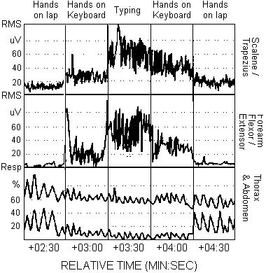
The following are common physiological patterns which may be identified and may increase risk (Fig. 4).


Figure 4. Common Dysfunctional Physiological Patterns include: Increased scalene/trapezius sEMG activity when the hands are on the keyboard and during typing. Increased forearm flexor/extensor sEMG activity when the hands are on the keyboard. Absence of micro-breaks (low sEMG epochs) when typing. Increased respiration rate when the hands are on the keyboard and when typing. Decreased abdominal expansion while breathing when hands are on the keyboard and when typing.
  • Absence of regenerative micro-breaks (1-2 seconds epochs of low sEMG activity every 10-20 seconds) from activated muscles.
  • Increased scalene/trapezius sEMG activity when the person's fingers are on the keyboard. (Covert dysponesis: the person is unaware the shoulders are raised when preparing to type.)
  • Increased forearm sEMG activity as long as the fingers are on the keyboard. (The sEMG increased even when the person thought the arms and hands were relaxed at the keyboard.)
  • Increased respiration rate and thoracic breathing when the person types (increased nervous arousal) and decreased respiratory sinus arrhythmia.
  • Increased sEMG activity, increased respiration rate and thoracic breathing after hands are placed back on lap (lack of or slow recovery).
  • Low correlation between subjective sense of stress/tension rating and sEMG activity.
PHASE 2:
Effect of Emotion on Physiology
PURPOSE:
To assess the impact of negative emotions upon physiology.

PROCEDURE:
  1. Sit comfortably in front of the computer, hands on lap.
  2. Think, feel, imagine, visualize an angry, resentful, and frustrating job-related or personal experience and indicate when these angry/resentful feelings/thoughts are present.
  3. Continue to experience the negative feelings/thoughts with hands on lap (30 seconds).
  4. Continue to experience the negative feelings/thoughts, and type a standard text (60 seconds).
  5. Let go of the negative feelings/thoughts and rest/relax with hands on lap (60 sec. baseline).
RISK PATTERNS:
The following are common risk patterns, in addition to those described in PHASE 1, which need to be retrained (Fig. 5).


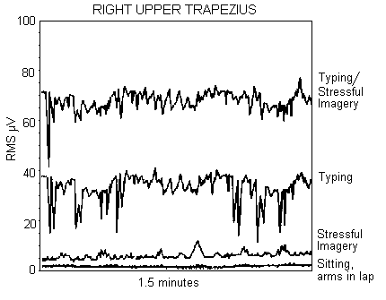
Figure 5. The effect of stressful imagery on right upper trapezius muscle activity. Note that there are NO micro-breaks during the typing with stressful imagery, while there are micro-breaks (epochs of low EMG) during the normal typing task. In addition, the EMG activity is significantly increased during stressful imagery in both the sitting, arms in lap, and the typing conditions.
  • Increased sEMG activity and arousal during typing as compared to PHASE 1 baseline and typing tasks.
  • Slow recovery back to baseline measures following the instructions of letting go of the negative imagery while resting hands on lap.
PHASE 3:
Effect of Continued Task Performance on Physiology
PURPOSE:
To assess the impact of long duration work pattern upon physiology.

PROCEDURE:
  1. Sit comfortably with hands resting (relaxed) on lap (1 minute baseline).
  2. Type a standard text (a sample letter or materials that simulate a normal job task) (10-50 minutes).
  3. Sit comfortably with hands resting (relaxed) on lap (1-5 minute baseline).
RISK PATTERNS:
The following are common risk patterns, in addition to those described in PHASE 1 and 2, which need to be retrained.

  • Sustained upper trapezius sEMG activity lasting longer than 30 seconds without the presence of regenerative micro-breaks (1-2 seconds of very low sEMG activity) (3).
  • Increased scalene/trapezius sEMG during data entry without micro-breaks.
  • Increased forearm sEMG while the fingers are on the keyboard.
  • Increased respiration rate and thoracic breathing during data entry.
  • Slow recovery back to baseline measures following typing task.
  • Absence of gross body movements.
PHASE 4:
Symmetrical Movement Analysis for Muscle Co-Contraction and Recovery Analysis
PURPOSE:
To assess sEMG imbalance during movement patterns (Taylor, 1993; Wilson, 1994; and Skubick, Clasby, Donaldson, & Marshall, 1993; Donaldson, 1994).

PROCEDURE For SCM sEMG assessment:
SENSOR PLACEMENTS:
Use two or four EMG channels, place sEMG active sensors on the right and left sternocleidomastoid (SCM) (optional: right and left upper trapezius)(Fig. 3).

  1. Sit comfortably in front of the computer with the hands on lap, while looking straight ahead (5 seconds).
  2. Rotate head to the right, as if looking over the right shoulder, while keeping the torso facing forward (5 seconds).
  3. Rotate head to face forward (5 seconds).
  4. Rotate head to the left, as if looking over the left shoulder, while keeping the torso facing forward (5 seconds).
  5. Rotate head to face forward (5 seconds).
  6. Repeat rotation sequence 5 times.
ALTERNATIVE MOVEMENT PATTERN:
Have person perform real job tasks such as reaching for the phone, manuals, or turning pages with one hand and then with the other hand. Repeat movement 5 times.

PROCEDURE For upper trapezius assessment:
SENSOR PLACEMENTS:
Use two or four EMG channels, place sEMG sensors on right and left upper trapezius (optional: right and left SCM).

  1. Sit comfortably in front of the computer with the arms and hands hanging along the sides of the body with the palms facing toward each other (5 seconds).
  2. While keeping the elbow straight, lift both arms up until they are horizontal (90 degrees to the body) and hold for 6 seconds. Then return the arm to hang along side the body. (In this movement, the palms initially point towards the floor.)
  3. Repeat movement sequence 5 times.
ALTERNATIVE MOVEMENT PATTERN:
Have person perform movements which mimic common job movements.

OPTIONAL:
Repeat one cycle of the above movement patterns while changing the time duration 30-60 seconds at the full extension. (Do not continue if pain occurs.) Allow at least 120 seconds for recovery between right and left movements.

RISK PATTERNS:
The following are common risk patterns, in addition to those described in PHASE 1, 2, and 3, which need to be retrained.

  • Asymmetry in sEMG activity during movement. For example, right SCM sEMG significantly higher when head turns left than left SCM when head turns right or vise versa; similarly, asymmetry in sEMG trapezius activity when arms are moved upward.
  • Significant co-contraction of antagonist during rotational movement. For example, left SCM sEMG is activated while head turns to the left.
  • Breath holding or very shallow breathing during movements.
  • Lack of awareness of breath holding.
  • Lack of awareness of co-contraction and asymmetrical muscle use patterns.
  • Slow sEMG recovery to baseline after five repetitions or after longer holding.
Data Review
Review the recorded data from the assessment protocol. Identify physiological response patterns and work habits which may increase the risk of RSI. The most commonly observed risk patterns are the absence of muscle tension awareness (awareness of muscle tension does not correlate with sEMG activity), inability to relax muscles, dysponesis (bracing of the trapezius during data entry), increased arousal during data entry, absence of regenerative micro-breaks during typing tasks (see Figs. 4 and 5), asymmetry and co-contractions during movement, and absence of gross body movement patterns.
Training and Education
The training protocol consists of reducing the observed risk patterns and generalizing these new skills into the persons work behavior. If significant dysponesis is observed, a more detailed sEMG analysis of specific muscles is required. The more specific the feedback, the more successful will be the skill acquisition.
The general training themes consist of increasing awareness of dysponetic activity, inhibiting co-contraction by tightening the correct agonist and inhibiting the antagonist, encouraging regenerative micro-epochs of very low sEMG activity of an activated muscle, reducing arousal (startle) during data entry through methods such as continued breathing, developing movement patterns using both sides of the body equally, and teaching that health consists of the alternation between activity and regeneration (movement and relaxation). The training goals are enhanced when monitored with portable EMG trainers (MyoTrac and MyoTrac2) or computer based systems (ProComp, FlexComp or MyoTrac2). The general psychophysiological concepts to achieve training goals are:
  • Encourage lowering of arousal during task performance and breaks.
  • Teach diaphragmatic breathing to reduce hyperventilation (Peper, 1990).
  • Teach momentary regenerative breaks during continued task performance. Sustained muscle activity of greater than 30 seconds needs to have regenerative epochs (1-2 seconds) of low EMG activity. The micro-breaks are much more important than the muscle tension during the task (Taylor, 1993).
  • Develop muscle strength, flexibility and bilateral symmetry appropriate for task performance through movement exercise and workstation rearrangement (Wilson, 1994).
CAUTION: Any numbness, tingling, prickling sensation or loss of sensation or dropping of objects should be evaluated by a physician. These symptoms are most prevalent in the early morning, evening or wakes the person from sleep.
TRAINING GOALS:
A. OPTIMIZE ERGONOMIC CORRECT POSITION
  1. Implement the ergonomic improvements derived from the ERGONOMIC WORK STYLE EVALUATION.
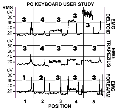
  2. Optimize body position at the work station with sEMG monitoring. For example, place sensors on deltoid or trapezius to identify the neutral arm position while hands are on keyboard (Fig. 6) (Peper and Shumay, 1994).

Figure 6. The use of sEMG from right trapezius, deltoid, and forearm to optimize keyboard location. Position 1 is resting with hands on lap, position 2 is most ergonomically correct with hands at keyboard, while position 3, 4, and 5 are ergonimacally incorrect. NOTE that there is NO correlation between sEMG activity and subjective awareness of tension as indicated by the numerical scores (1 - most relaxed to 5 - most tense) for each position and shoulder and forearm body locations.
B. EMG GOALS
  1. Inhibit scalene/trapezius sEMG activity while fingers are on the keyboard during rest and data entry. This means the person learns to sense bracing in the shoulders and lets the shoulders stay relaxed during data entry.
  2. Inhibit finger/wrist flexor/extensor sEMG activity when fingers are resting on keyboard.
  3. Inhibit shoulder girdle and arm bracing (excessive sEMG activity) while using a mouse.
  4. Inhibit sEMG co-contraction of muscles such as SCM.
  5. Teach scapular stabilization utilizing lower trapezius and serratus anterior sEMG feedback to inhibit upper trapezius activity (Bender, 1993).
  6. Monitor sEMG and inhibit dysponetic activity from relevant muscle groups while performing job related keyboard entry tasks.
C. RELAXATION/STRENGTHENING PRACTICES
  1. Head rotations: SLOWLY look over right shoulder. Hold 20 seconds, back to center. Repeat on the other side. (Minimize shoulder movement as much as possible.)
  2. Side headbends: Put right ear to right shoulder. Hold 20 seconds, back to center. Repeat on other side (minimize shoulder movement as much as possible).
  3. Turkey pull: GENTLY pull your neck backwards as if someone had a string attached to the back of your neck and was pulling it backward. Keep the jaw parallel to the ground and shoulders relaxed. Do 2-30 times daily.
  4. Shrug shoulders backward and forward in a circular motion, go slowly. Several circles should be executed-- each of a different diameter.
  5. Place arms at sides as if you were standing at attention. Keeping arms as straight as possible, raise them up over your head until the backs of your hands meet above your head. Ensure that palms face down as arms extend. The action should look like a slow motion jumping jack (or a very lethargic duck trying to fly). Do not arch lower back.
  6. Do Dynamic Relaxation of the neck, shoulders, arms, wrists, hands, and fingers (Peper and Holt, 1993). Teach internal mastery of high and low muscle tension and the ability to relax the muscle at will.
  7. Take brief 1 to 2 seconds regenerative breaks every 30 seconds during keyboard data entry and mouse use. For example, drop hands to the desk top or lap, the sEMG of the neck and shoulders should instantly return to low baseline levels.
D. EMOTIONAL CONTROL
  1. Enhance awareness of how negative emotions contribute to dysfunctional patterns.
  2. Develop communication and problem solving skills to resolve work and/or family conflicts.
  3. Teach thought stopping and/or task focusing exercises.
E. IMPLEMENTATION
  1. Generalize the above learned skills while performing relevant keyboard entry tasks at the actual job site.
  2. Breathe diaphragmatically and decrease breathing rate during relevant task performance.
Suggestions and Implications
Every person who uses or begins to use a computer should be instructed in somatic awareness, proper ergonomics, and rest/activity cycles. For many people, a signal from a small portable sEMG feedback device can help facilitate awareness of dysponesis during data entry and mouse use. Both the MyoTrac and MyoTrac2 devices (from Thought Technology Ltd.), offer the option for delayed tone feedback. This delayed feedback ignores the normal stretching, yawning, and other brief movements, whereas sustained sEMG activity triggers a warning feedback tone. In addition, external reminders to trigger brief regenerative breaks, as well as, encouraging episodic body movements may reduce the risk of RSI (e.g., timed alarms or automatic data entry interrupts). Finally, this protocol can be used to teach computer users preventative skills to avoid RSI and mobilize health. Table 1:
TYPICAL COMPUTER USER COMPLAINTS, CORRESPONDING ERGONOMIC FACTORS AND SUGGESTED MUSCLE GROUPS FOR SENSOR PLACEMENT
HEAD

Typical complaints Possible ergonomic factors Areas of sEMG placement
Eye strain:
Pain or fatigue
VDT distance and tilt
Lighting
Optical prescription
Frontalis/posterior neck
Temporalis
Suboccipital
Frontal
Headache Telephone usage
Rest breaks from VDT
Sitting posture
Squinting
Clenching teeth
Fumes of toner
Fluorescent lights
Frontal/posterior neck
Temporalis
Masseter
Suboccipital
Cervical paraspinal
Trapezius
Sternocleidomastoid
NECK AND SHOULDERS

Typical complaints Possible ergonomic factors Areas of sEMG placement
Headache
back of head or neck
Document position (flat)
Distance from VDT (too far)
Head off-center
Arms not supported
Bifocals
Frontal/posterior neck
Trapezius
Masseter
Sternocleidomastoid
Pain in shoulders Head tilt (display too high
or too low
Bifocal glasses
Hunched shoulders (keyboard
height too high)
Drooped shoulders (too far
away from keyboard.
no muscle tone)
Mouse distance
One-sided movements
Trapezius
Deltoid
Pectoralis
Infraspinatus
Scalenes
ARMS, WRISTS AND FINGERS

Typical complaints Possible ergonomic factors Areas of sEMG placement
Elbow pain Repetitive motion/no rest
Keyboard design
Mouse distance
Triceps
Brachioradialis
Forearm extensors
Scalenes
Wrist pain Repetitive motion/no rest Upper shoulder
Sternocleidomastoid
Brachioradialis
Forearm extensors
Scalenes
Finger pain Repetitive motion/no rest Digital Extensors
Forearm extensors
Scalenes
BACK

Typical complaints Possible ergonomic factors Areas of sEMG placement
Lower back or hip pain Chair (lumbar support)
Posture
Leg Position
Feet on floor (sloped rest)
High/low hip or shoulders
Hip beneath knee
Upper back
Lumbar region
LEGS, ANKLES AND FEET

Typical complaints Possible ergonomic factors Areas of sEMG placement
Pain down the leg Compression from chair
(inadequate padding on bottom,
sharp front edge)
Leg position (crossed legs
or sharp leg bend
One side movement
Hamstrings
Glutei
Piriformis
Swollen ankles Lack of movement
Pain in foot or toes Feet not on floor/sloped rest
Poor circulation
Leg position
Lack of movement


SFSU WORKSTATION AND ERGONOMIC ASSESSMENT
(Peper & Tibbets, 1994)

Instructions: Observe and circle possible risk factors.
Name: ____________________ Date: _____________ Rater: ____________________


Monitor     80 degrees from eye position      High ___   Low ___
              Distance 3 feet                          Near ___   Far ___   ? ___
              Position                                    Left ___ Right ___   ? ___
              Glare and reflexion                      Yes  ___     ? ___
              Bright lights                              Yes  ___     ? ___
              Needs larger screen                  Yes  ___     ? ___
              High.clear screen resolution       No   ___     ? ___
              Ability to look at a far object       No   ___     ? ___
              Glasses-bifocals                       Yes  ___     ? ___
              Contacts                                  Yes  ___     ? ___
              Uses reading/computer glasses    Yes  ___     ? ___
              Position of reading material        Near ___ Far   ___
                                                             High ___ Low   ___

Comments:________________________________________________________________

Chair        Feet reach floor                          No   ___     ? ___
              Thighs compressed                    Yes  ___     ? ___
              Seat angles back                       Yes  ___     ? ___
              Back support                             No   ___
              If present, is it used?                  No   ___     ? ___
                                                              High ___   Low ___
              Arm rests                                  Yes  ___
              Lifts shoulders                           Yes  ___     ? ___

Comments:________________________________________________________________


Legs         Enough room for feet                  No   ___     ? ___
              Space for leg movement              No   ___     ? ___
              Angle of knees at 110o                No   ___     ? ___
              Foot support                               No   ___     ? ___
              If present, is it used                    Yes  ___    No ___
                                                               Sometimes     ___

Comments:________________________________________________________________


Keyboard   Adjustable                     No   ___     ? ___
                                                   High ___   Low ___
               Stable                           No   ___     ? ___
               Wrist pad                      No   ___     ? ___
                                                 Hard ___     ? ___

Comments: _____________________________________________________________


Arms      Angle of upper arm (vertical)       Forward ___ Backward ___
              Angle of forearm at elbow (110o)  Smaller ___ Greater  ___
              Elbows out                               Yes  ___        ? ___
              Angle of hand                            Flexed ___ Extended ___
              Finger nails                               Long ___        ? ___
              Types with finger nails                 Yes ___        ? ___
              Tension visible (tendons, fingers)    Yes ___        ? ___

Comments: _____________________________________________________________


Mouse/        Present                           Yes ___        ? ___
trackball       Frequency of use              Often  ___ Sometimes___
                   Location                        Distant ___        ? ___
                   Reaches out to side           Yes ___        ? ___
                   Reaches out forward           Yes ___        ? ___

Comments: _____________________________________________________________


Phone         Frequency of use                    Often  ___ Sometimes___
                  Duration of use                        ___ Minutes
                  Twists for use                         Yes ___        ? ___
                  Location                                 Distant ___
                  Shoulder rest                            No ___        ? ___
                  Headset                                  No ___        ? ___
                  Writing.data entry during calls   Yes ___        ? ___

Comments: ___________________________________________________________


Space         Enough room on desk                No ___        ? ___
                  Office space personalized           No ___        ? ___
                  Back to door                              Yes ___      ? ___

Comments: _____________________________________________________________

Work habits   Takes breaks every 15-30 min.            No ___       ? ___
                     Does multiple body movements/task   No ___       ? ___
                     Assymetrical movements during task  Yes ___      ? ___
                     Reaching during work                       Yes ___      ? ___
                     Mini-breaks during keyboard entry       No ___        ? ___
                     Percent time at computer                 <25% 50% 75%>  ? ___
                     Previous workstation assessment
                     and/or suggestions implemented?        No ___        ? ___

Comments: ____________________________________________________________


Personal      Clothing                                         tight ___      ? ___
                   Belt (waist constricted)                   Yes ___      ? ___
                   Stationary (abscence of movement)   Yes ___      ? ___
                   Thoracic breath pattern                    Yes ___      ? ___
                   Shifts to thoracic breath
                   pattern while working                       Yes ___      ? ___
                   Skewed posture while sitting              Yes ___      ? ___
                   Startle posture                                 Yes ___      ? ___
                   Conscientious/driven                        Yes ___      ? ___
                   Hands                                           Cold ___  Moist ___      ? ___
                   Smoking                                        Yes ___      ? ___

Comments: ___________________________________________________________

Workstation adjustment suggested: ________________________________________

__________________________________________________________________________

__________________________________________________________________________

__________________________________________________________________________

Changes in work habits  suggested: _______________________________________

__________________________________________________________________________

__________________________________________________________________________

__________________________________________________________________________


Psychophysiological profile appointment date: _________________________________


RECOMMENDED SOURCES
  1. Basmajian, J.V. & Blumenstein, R. (1980). Electrode Placement for EMG Biofeedback. Baltimore: Williams & Wilkins.
  2. Cram, J. (Ed). (1989). Clinical EMG for Surface Recording. Nevada City, CA: Clinical Resources.
    Grandjean, E. (1987). Ergonimics in Computerized Offices. New York: Taylor and Francis.
  3. Peper, E. (1990) Breathing for Health. Montreal: Thought Technology Ltd.Soderberg, G.L. (Ed). (1992).
  4. Selected Topics in Surface Electromyography for use in the Occupational Setting: Expert Perspectives. U.S. National Institute for Occupational Safety and Health, Publication No. 91-100. Available from NIOSH Publications, Mail Stop C-13, 4676 Columbia Parkway, Cincinnati, Ohio 45226-1992. State of California, Department of Personnel Administration. (1993). Video Display Terminal (VDT) Users Handbook. Sacramento, CA.
  5. Travell, J.G. & Simons, D.G. (1983). Myofascial Pain and Dysfunction: The Trigger Point Manual. Baltimore: Williams & Wilkins.
  6. Whatmore, G. and Kohli, D. (1974). The Physiopathology and Treatment of Functional Disorders. New York: Grune and Stratton, 1974.
Footnotes:
  1. A number of the concepts in this protocol have been derived and expanded from the contributions made by Donaldson & Skubick, 1993; Ettare & Nadler, 1993; and Middaugh et al, 1993. We thank Cathy Holt, Dianne Shumay, and Scott Davis for their helpful contributions.
  2. Reprint requests contact: Erik Peper, Ph.D., Institute for Holistic Healing Studies, San Francisco State University, 1600 Holloway Ave, San Francisco, CA 94132. FAX: 415-338-0573; EMAIL: epeper@sfsu.edu.
  3. Ergostats by Will Taylor, M.D., software to analyze sEMG recordings for regenerative micro breaks is available from biofeedback distributors.